
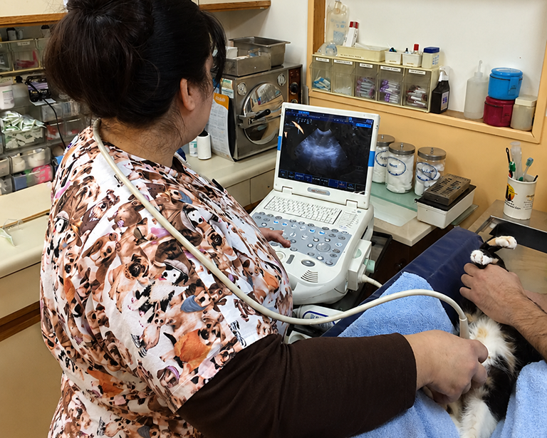
Detailed Ultrasound Imaging for Soft Tissues
Ultrasonography provides real-time views of internal organs, blood flow, and soft tissue structures invisible on X-rays. This non-invasive technology helps us evaluate the heart, abdomen, reproductive system, and pregnancy with remarkable detail.
Performed without anesthesia in most cases, ultrasound is comfortable, painless, and immediately informative for our veterinary team. We use it to guide biopsies, confirm pregnancies, assess organ disease, and monitor treatment responses over time.
- Visualizes organ texture, fluid accumulation, masses, and structural changes radiographs cannot reveal.
- Guides minimally invasive biopsies and fluid aspirations with pinpoint accuracy and safety.
- Pregnancy confirmation and litter monitoring available beginning approximately twenty-eight days post-breeding.
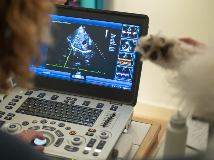
- Cardiac ultrasound evaluates heart function, valve integrity, and circulation patterns in detail.
Frequently asked Questions
Curious about how ultrasonography works and what your pet experiences during scanning? These commonly asked questions explain the procedure, preparation requirements, and diagnostic capabilities of ultrasound imaging.
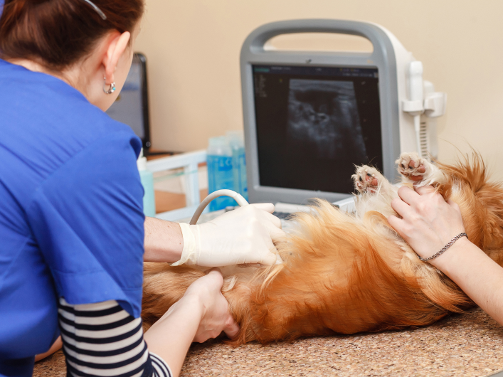
Yes, the area being imaged must be shaved because hair traps air and blocks sound waves from producing clear diagnostic pictures of internal structures.
Ultrasound visualizes soft tissue detail and movement in real-time, while radiographs show bone, gas, and overall organ silhouettes in static images.
Ultrasound itself is painless, though pets sometimes find lying still on their back for fifteen to thirty minutes mildly uncomfortable or unusual.